
SUCCESSFUL STORIES
Computer Aided Diagnosis
Worked with one of the largest hospitals in China, we have developed a medical image based CAD system for lung cancer. Lung cancer is one of the leading causes for cancer related mortality around the world. Near 180,000 new lung cancer cases are diagnosed each year in US. High mortality of lung cancer, of a rate about 90% , contribute to about 29% of total cancer caused mortality.
Successful early detection can greatly improve the patients’ survival rate. If diagnosed and treated at an early-stage, lung cancer patients’ five-year survival rate can be improved dramatically from about 13% to 41%. However, making a confirmative diagnosis for all pulmonary nodules\masses is still an open issue, particularly for current trends to screen lung cancer using low-dose CT scans. Moreover, overwhelming image data generated by existing imaging tools make manual screening impossible.
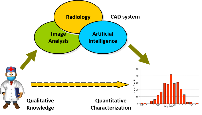
- Computer aided diagnosis (CAD). CAD is a cross-disciplinary research area covering radiology, image processing and analysis, artificial intelligence and machine learning, nonlinear analysis, and nonlinear feature extraction and selection. In this project, we aimed to
- Develop a system for medical image browsing, manipulating, and enhancement;
- Develop an efficient system for automatic nodule screening to help early detection of lung cancers integrating image analysis and machine learning technologies.
- Develop an effective system for diagnosis of nodules and other abnormalities with image processing, nonlinear analysis, and machine learning technologies.
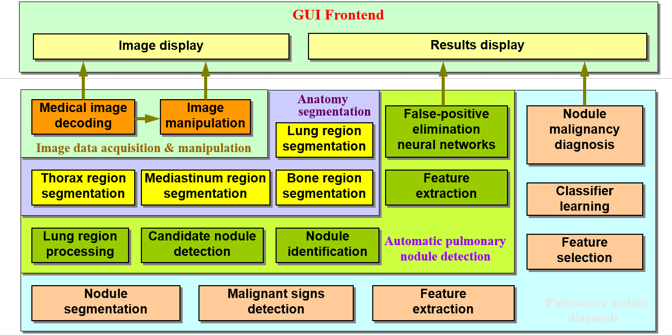
Our system is composed of four main modules with following functions: image data acquisition and manipulation, anatomy segmentation, automatic pulmonary nodule detection, and pulmonary nodule diagnosis. Each module has several sub-modules. Our proprietary TRUST-TECH methodology had been applied to solve the involved optimization problems in realizing this system, covering the tasks of optimal object detection, image segmentation, and optimal design of classifiers for the diagnosis.
Diagnosis performance is evaluated in two ways: one way is evaluated by image slices, the other by patient. The performance by patient is obtained based on the results by slice through a committee machine technique.
| Evaluation Method | Total | Right | False |
| By Slice | 442 slices | 355 (80.3%) | 87 (19.7%) |
| By Patient | 74 patients | 52 (70.3%) | 22 (29.7%) |